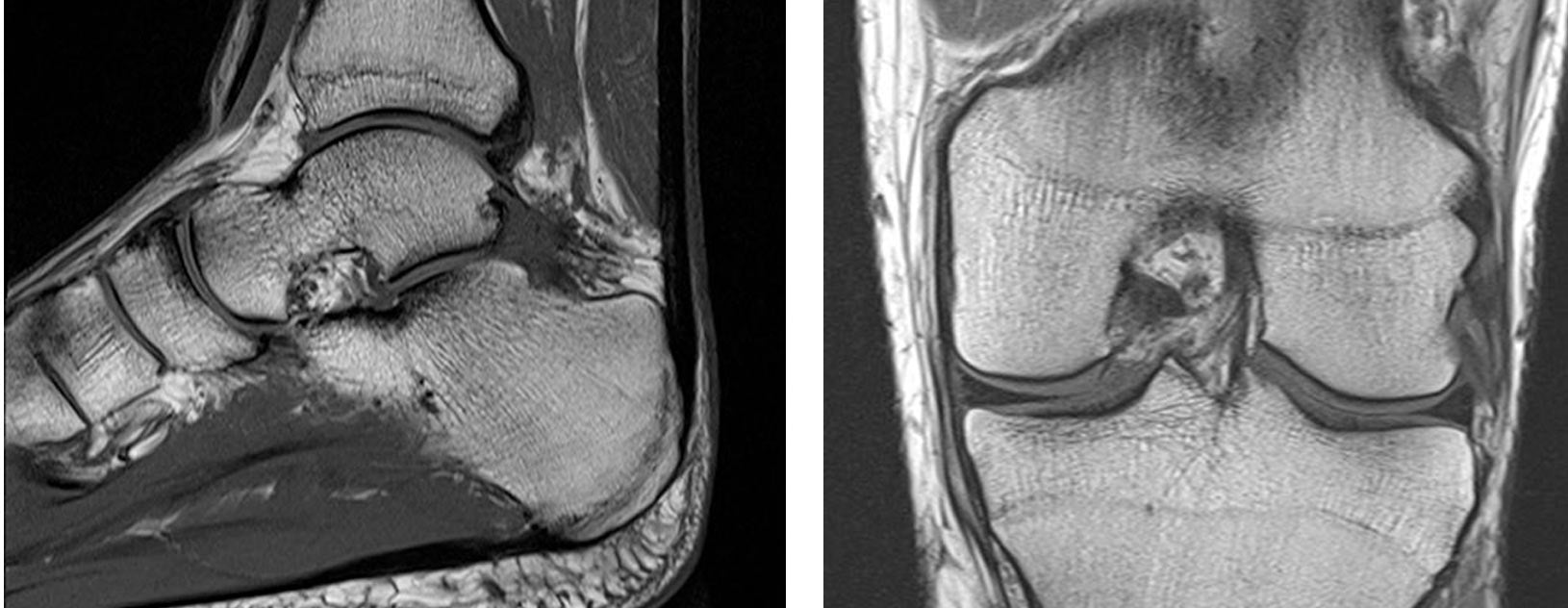
MRI
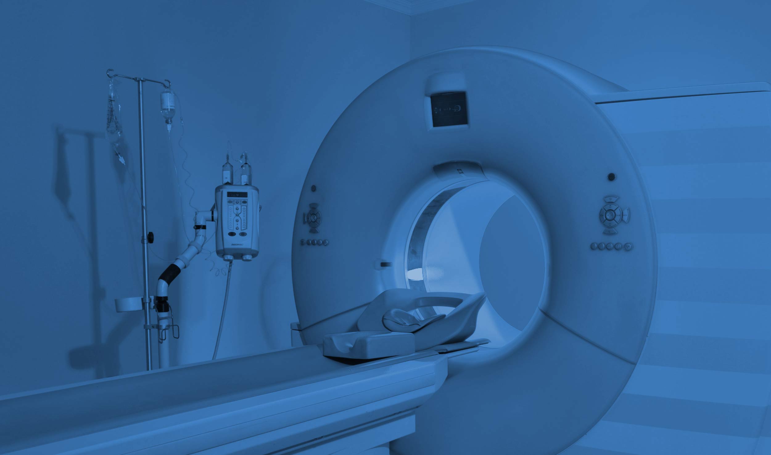
Magnetic Resonance Imaging is a non-invasive technique which uses a combination of magnetism and radio waves to produce a detailed series of images of the body part concerned. It does not involve radiation and is an extremely safe form of imaging. Depending on the body part being imaged the patient may have to partly of fully enter the magnet but it will not come into contact with the patient at any point.
MRI Arthrography involves MRI imaging of a large joint such as shoulder, hip, wrist and elbow following injection of “dye” into the joint. The radiologist performs this injection into the joint using a needle under ultrasound or fluoroscopic guidance.
Owing to the nature of the magnet used in MRI scanners there are some contra indications which mean it may not be suitable for people with the following:
- Pacemakers – although some of these may now be compatible with the MRI scanner
- Cochlear implant
- Metallic fragments in the eyes from previous injury
- Spinal stimulator devices
- Cardiac valves/stents – some of these are safe and the exact details of the implant will be needed to ensure the scan can go ahead
For safety reasons a questionnaire is used prior to all examinations in order to allow the scans to go ahead safely. Please feel free to ask the department if you have any doubts.
Our team of musculoskeletal radiologists will send a report back to the referring clinician explaining the imaging findings and any further recommendations.